
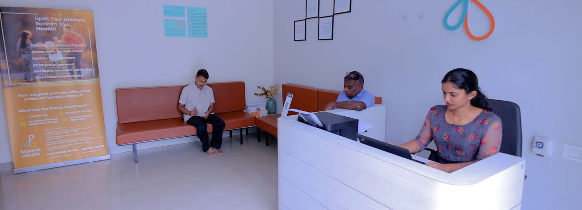
Maana Spine, Joint & Wellness Clinic at Aluva
Welcome to Maana Spine, Joint & Wellness Clinic, Aluva’s premier destination for advanced, non-surgical spine and joint care. Located near Pulinchud Metro Station, Kunnathery Thikavu, our state-of-the-art wellness center offers comprehensive treatment solutions for chronic back pain, neck pain, joint conditions, along with innovative medical weight managementand sports rehabilitation services. As Aluva’s leading spine clinic and joint wellness clinic, we combine FDA-approved technology with expert physiotherapy to deliver exceptional results without surgery.
Our spine, joint & wellness clinic in Aluva has helped patients avoid surgery through our comprehensive, evidence-based treatment programs. Whether you’re suffering from chronic back pain, disc problems, joint discomfort, seeking sustainable weight loss, or recovering from sports injuries, our joint wellness clinic in Aluva provides personalized, evidence-based care that addresses the root cause of your condition.
Advanced Spine & Joint Treatment in Kunnathery Thikavu, AluvaWhy Choose Our Spine Clinic in Aluva?
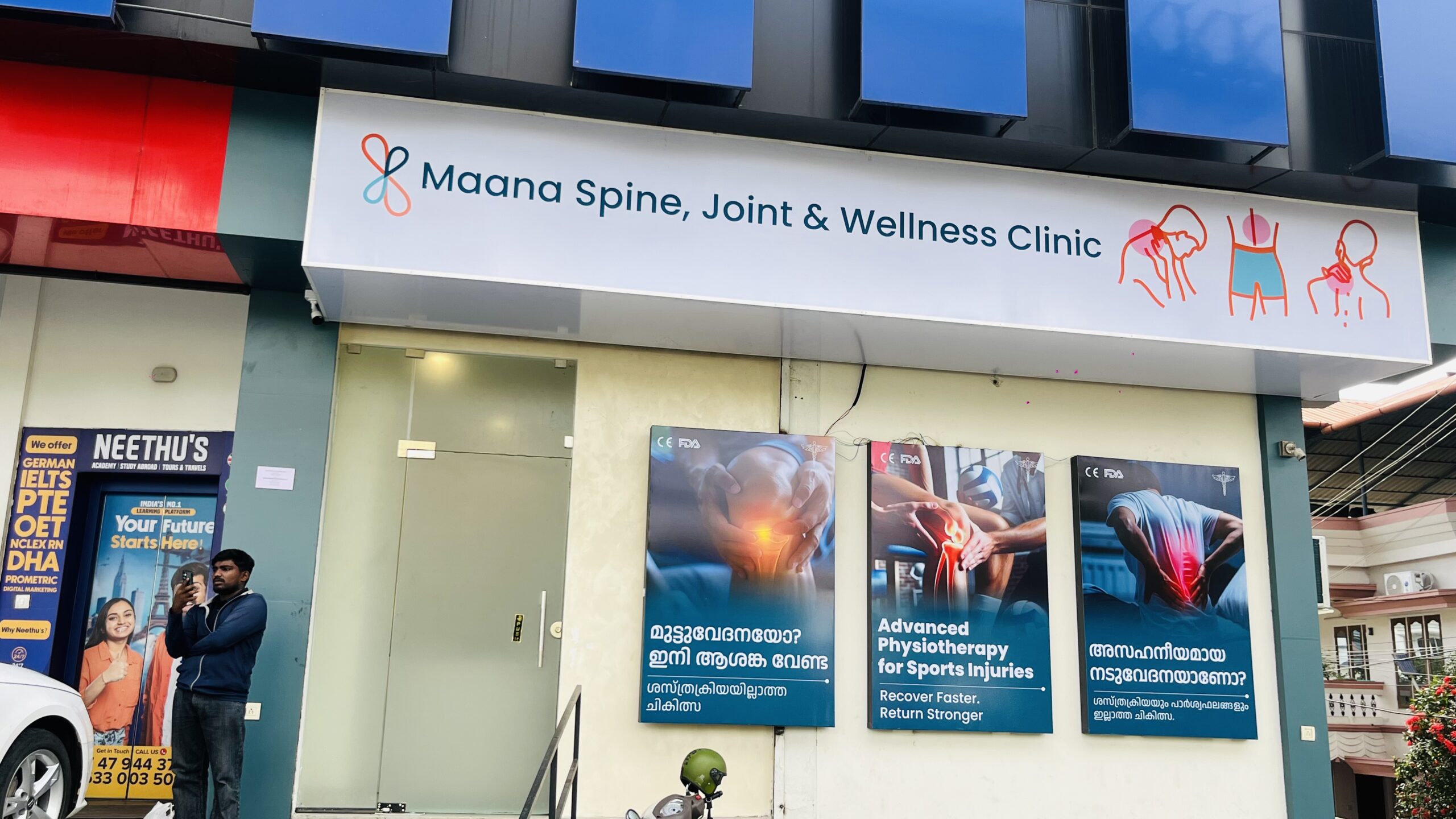
At Maana Spine, Joint & Wellness Clinic, we revolutionize back pain treatment in Aluva through cutting-edge, non-surgical methods. Our clinic stands as the only comprehensive spine, joint & wellness center in Aluva, offering:
- FDA-Approved & CE-Approved Spine Technology: US & European-standard spinal decompression systems
- Expert Medical Team: Board-certified specialists in non-surgical spine and joint care
- Proven Results: A patient-centered, non-surgical approach with lasting pain relief
- Convenient Location: Easily accessible at Kunnathery Thikavu, serving all of Aluva
- Comprehensive Care: Complete spine, joint, wellness, and weight management solutions
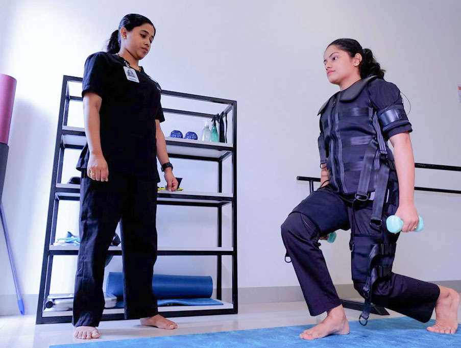
Our Core Specializations
Non-Surgical Spine Treatment in Aluva
Our non-surgical spine treatment in Aluva utilizes advanced spinal decompression therapy, eliminating the need for invasive procedures. This revolutionary treatment effectively addresses:
- Chronic lower back pain and neck pain
- Disc bulge and herniation without surgery
- Sciatica and nerve compression relief
- Spinal stenosis and degenerative disc disease
- Work-related spine injuries and postural problems
Advanced Joint Wellness Programs
As Aluva’s premier joint wellness clinic, we offer comprehensive joint care including:
- Joint pain treatment in Aluva for knee, shoulder, and hip conditions
- Advanced physiotherapy for joint mobility restoration
- Arthritis management and non-surgical joint preservation
- Post-surgical joint recovery and rehabilitation
- Age-related joint problems and mobility enhancement
Comprehensive Physiotherapy Services in Kunnathery Thikavu
Our physiotherapy clinic in Kunnathery Thikavu features state-of-the-art treatment modalities:
- Advanced Physiotherapy in Aluva: Manual therapy and exercise rehabilitation
- Pain Management: Multi-modal approaches for chronic conditions
- Movement Restoration: Functional rehabilitation and mobility training
- Ergonomic Training: Workplace injury prevention and posture correction
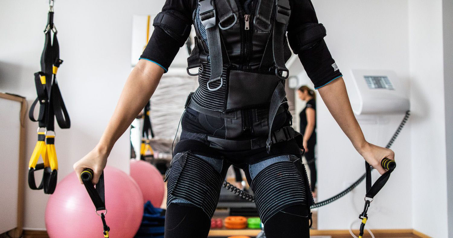
Medical Weight Management Program – Revolutionary in Kerala
Kerala’s First EMS Training + Behavioral Therapy Weight Loss Solution
Introducing our groundbreaking medical weight management program in Aluva!
Our evidence-based medical weight loss program in Aluva combines cutting-edge technology with psychological support:
EMS Training for Weight Loss – First in Kerala
- Revolutionary Technology: Electrical muscle stimulation burns up to 500 calories in 20 minutes
- Muscle Building + Fat Loss: Simultaneous muscle strengthening and weight reduction
- Time-Efficient: Equivalent to 4 hours of traditional exercise in just 20 minutes
- Medical Supervision: Safe, monitored sessions by qualified professionals
Acceptance-Based Behavioral Therapy for Weight Management
- Evidence-Based Psychology: Clinically proven approach for sustainable weight loss
- Mindset Transformation: Address emotional eating and lifestyle patterns
- Long-term Success: Build healthy relationships with food and exercise
- Professional Support: Licensed therapists specializing in weight management
Medical Weight Management Results
- 12-15% Body Weight Loss: Achieve significant, measurable results safely
- Doctor Supervised: Medical monitoring throughout the program
- Sustainable Approach: Long-term lifestyle changes, not quick fixes
- Comprehensive Care: Nutrition, exercise, and psychological support integration
Sports Rehabilitation & Musculoskeletal Therapy Services
Advanced Sports Medicine & Physical Rehabilitation
Building on our spine and joint expertise, we offer comprehensive sports rehabilitation services:
Sports Injury Rehabilitation in Aluva
- Athletic Injury Recovery: ACL/MCL tears, rotator cuff injuries, sports trauma
- Overuse Injury Treatment: Tennis elbow, runner’s knee, swimmer’s shoulder
- Return-to-Sport Programs: Safe, progressive activity resumption protocols
- Performance Enhancement: Athletic conditioning and injury prevention
Musculoskeletal Rehabilitation Services
- Workplace Injury Recovery: Repetitive strain injuries and occupational health
- Chronic Pain Management: Multi-disciplinary approach to persistent conditions
- Post-Surgical Rehabilitation: Comprehensive orthopedic recovery programs
- Movement Disorders: Biomechanical assessment and correction
Comprehensive Wellness Services in Kunnathery Thikavu
State-of-the-Art Treatment Facility in Kunnathery Thikavu
Our physiotherapy clinic in Kunnathery Thikavu features the most advanced treatment modalities available in Kerala:
Advanced Treatment Technologies
- Spinal Decompression Systems: Computer-controlled decompression for disc problems
- EMS Training Equipment: Revolutionary muscle stimulation for fitness and weight loss
- Manual Therapy Stations: Expert hands-on treatment techniques
- Exercise Rehabilitation Areas: Customized strengthening and conditioning programs
- ESWT & Cryotherapy Facilities: Advanced modalities for rehabilitation and recovery
Specialized Treatment Programs
Our advanced physiotherapy in Aluva includes specialized programs for:
- Spine & Joint Patients: Chronic pain relief and mobility restoration
- Weight Management Clients: EMS training and behavioral therapy integration
- Corporate Professionals: Workplace injury prevention and wellness programs
- Athletes & Active Individuals: Sports injury recovery and performance enhancement
- Senior Citizens: Age-related mobility and joint health programs
Our Comprehensive Treatment Approach
The Maana Difference: Integrated Healthcare Solutions
What sets our spine clinic in Aluva apart is our comprehensive, patient-centered approach:
Detailed Assessment & Diagnosis
- Spine & Joint Evaluation: Advanced imaging analysis and physical examination
- Weight Management Assessment: Body composition analysis and metabolic evaluation
- Movement Analysis: Biomechanical assessment for sports and daily activities
- Lifestyle Assessment: Comprehensive evaluation of occupational and activity factors
- Personalized Treatment Planning: Customized protocols across all services
Advanced Treatment Modalities
- FDA-Approved & CE-Approved Spinal Decompression: Non-surgical disc and nerve treatment
- Joint Restoration Therapy: Advanced joint care and mobility techniques
- EMS Training Technology: Revolutionary muscle stimulation for multiple applications
- Behavioral Therapy: Professional psychological support for sustainable changes
- Pain Management: Multi-disciplinary approaches for comprehensive relief
Long-term Wellness Support
- Ongoing Monitoring: Regular progress assessment across all programs
- Lifestyle Modification: Comprehensive guidance for lasting health changes
- Prevention Strategies: Injury and condition recurrence prevention
- Maintenance ProgramsMaintenance Programs: Sustained wellness and health optimization
Why Patients Choose Our Aluva Clinic
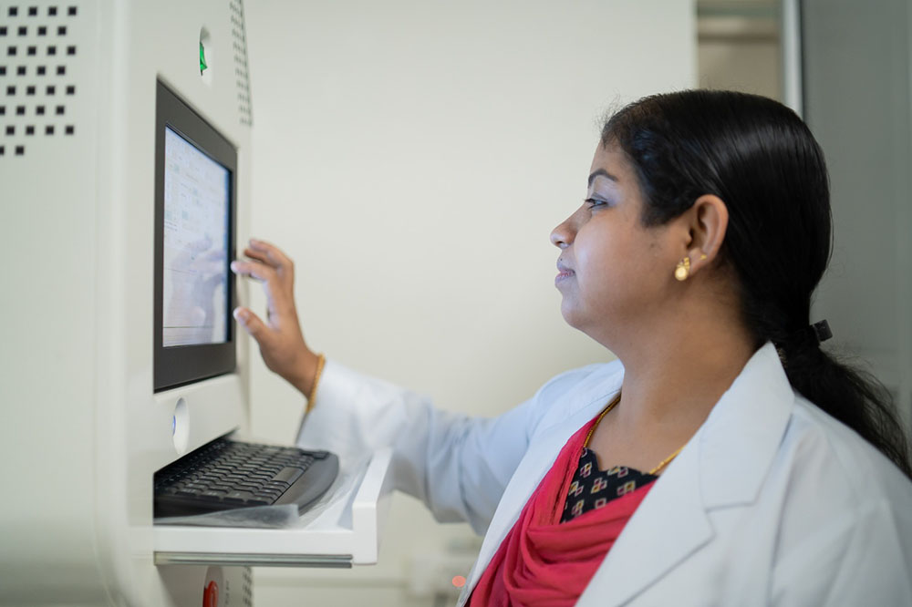
Advanced Technology & Equipment
Our wellness center in Aluva features:
- Computer-controlled spinal decompression systems for spine care
- State-of-the-art EMS training equipment for weight management
- Advanced physiotherapy equipment for joint rehabilitation
- Modern diagnostic tools and body composition analyzers
- Comfortable, private treatment environments for all services
Complete Services Offered at Our Aluva Location
Priority 1: Spine Care Excellence
- Chronic Back Pain Treatment: Comprehensive non-surgical solutions for lasting relief
- Neck Pain Therapy: Advanced cervical spine care and rehabilitation
- Disc Problem Management: Non-surgical disc bulge and herniation treatment
- Sciatica Relief: Targeted nerve decompression and pain management
- Spinal Stenosis Care: Advanced treatment options without surgery
- Postural Correction: Workplace and lifestyle-related spine issues
Priority 2: Joint Wellness Programs
- Knee Pain Management: Comprehensive knee care and arthritis treatment
- Shoulder Rehabilitation: Expert shoulder joint treatment and mobility restoration
- Hip Joint Care: Advanced hip mobility and pain management programs
- Arthritis Management: Holistic joint preservation and pain relief strategies
Joint Mobility Enhancement: Range of motion and functional improvement
Priority 3: Medical Weight Management Services
- EMS Training Programs: Revolutionary 20-minute effective workout sessions
- Behavioral Therapy Sessions: Professional psychological support for weight loss
- Medical Supervision: Doctor-monitored safe weight loss protocols
- Nutrition Counseling: Evidence-based dietary guidance and meal planning
- Body Composition Analysis: Advanced monitoring and progress tracking
- Lifestyle Coaching: Comprehensive support for sustainable changes
Priority 4: Sports Rehabilitation & Musculoskeletal Care
- Sports Injury Recovery: Comprehensive rehabilitation for athletic injuries
- Workplace Injury Treatment: Occupational health and repetitive strain recovery
- Movement Analysis: Biomechanical assessment and correction
- Performance Enhancement: Athletic conditioning and injury prevention
- Return-to-Activity Programs: Safe, progressive resumption protocols
Wellness & Prevention Services
Corporate Wellness Programs
Comprehensive workplace health solutions
Ergonomic Assessments
Workplace injury and postural problem prevention
Health Education
Patient empowerment through comprehensive knowledge
Maintenance Programs
Long-term wellness across all service areas
Convenient Location in Kunnathery Thikavu, Aluva
Easy Access from Across Aluva
Our physiotherapy clinic in Kunnathery Thikavu is strategically located for easy access from all parts of Aluva:
Address:
Near Pulinchud Metro Station, Kunnathery Thikavu
Aluva, Kerala 683106
Serving Patients From
- Kunnathery Thikavu & Surrounding Areas: Walking distance convenience for spine and joint care
- Central Aluva: Easy accessibility for weight management programs
- Ernakulam District: Well-connected for comprehensive wellness services
- Kochi Metro Line 1 Corridor: Quick commute for patients travelling by metro
Nearby Landmarks
- Steps from Pulinchodu Metro Station (Kochi Metro Line 1)
- About 1 km from Aluva Railway Station
- Close to Erumathala, Pallikkunnu, and SN Puram
Meet Our Expert Team
Qualified Medical Professionals
Our spine clinic in Aluva is staffed by highly qualified professionals:
- Board-Certified Physicians: Specialists in non-surgical spine and joint care
- Licensed Physiotherapists: Advanced training in spine, joint, and wellness therapy
- Weight Management Specialists: Medical doctors and behavioral therapists
- Rehabilitation Experts: Specialists in movement restoration and injury recovery
- Wellness Coordinators: Dedicated patient support across all programs
Continuing Education & Excellence
Our team regularly updates their skills through:
- International training in spine care and weight management technologies
- Advanced certification in EMS training and behavioral therapy
- Research and development in evidence-based treatment protocols
- Best practice implementation across all service areas
Treatment Programs & Packages
Comprehensive Treatment Options
18-Day Intensive Spine Program
Our signature back pain treatment in Aluva includes:
- Daily spinal decompression and physiotherapy sessions
- Progressive spine rehabilitation protocols
- Lifestyle modification and ergonomic guidance
- Nutritional support and follow-up care plans
Joint Wellness Rehabilitation Programs
Comprehensive joint pain treatment in Aluva:
- Customized joint mobility and strengthening programs
- Arthritis management and pain relief protocols
- Movement restoration and functional improvement
- Long-term joint health maintenance strategies
6-Month Medical Weight Management Program
Revolutionary weight loss program combining:
- 2 EMS training sessions per week for muscle building and fat loss
- Weekly behavioral therapy sessions for mindset transformation
- Medical monitoring and body composition tracking
- Nutrition counseling and comprehensive lifestyle coaching
- Achieve 12-15% body weight loss safely and sustainably
Corporate Wellness Packages
Specialized programs for Aluva businesses:
- On-site ergonomic assessments for spine and joint health
- Employee wellness workshops covering weight management
- Group treatment packages for workplace injury prevention
- Executive health programs integrating all services
Maintenance & Prevention Plans
Long-term wellness support:
- Monthly follow-up sessions for spine, joint, and weight management
- Exercise and nutrition program updates across all areas
- Lifestyle coaching and comprehensive health monitoring
- Preventive care strategies for sustained wellness
Frequently Asked Questions
What makes your spine clinic different from others in Aluva?
Our spine, joint & wellness clinic in Aluva is unique because we offer FDA-Approved & CE-approved spine technology PLUS Kerala’s first medical weight management program with EMS training and behavioral therapy. We’re the only clinic providing comprehensive spine, joint, weight, and wellness care under one roof with proven results.
How effective is non-surgical spine treatment?
Our non-surgical spine treatment in Aluva has helped patients avoid surgery while achieving lasting pain relief. Research supports non-surgical decompression as an effective option for many spine conditions when treated with our advanced spinal decompression protocols.
What conditions do you treat at your joint wellness clinic?
Our joint wellness clinic in Aluva treats osteoarthritis, sports injuries, post-surgical rehabilitation, chronic joint pain, mobility issues, and age-related joint problems using advanced, non-invasive techniques.
How effective is the EMS training for weight loss?
Our EMS training weight loss program in Aluva helps patients lose 12-15% of their body weight when combined with behavioral therapy. EMS sessions burn up to 500 calories in 20 minutes – equivalent to 4 hours of traditional exercise.
What is acceptance-based behavioral therapy for weight loss?
Our behavioral therapy weight loss approach focuses on changing your relationship with food and exercise through evidence-based psychological techniques. It addresses emotional eating, builds sustainable habits, and ensures long-term weight management success.
Do you accept insurance for physiotherapy treatments in Kunnathery Thikavu?
Yes, our physiotherapy clinic in Kunnathery Thikavu is reimbursed by major health insurance plans for spine and joint treatments. However, cash-less reimbursement facilities are not provided by most insurance companies for outpatient services in India. We also offer flexible payment options and corporate packages for weight management and wellness programs.
What non-surgical treatments are available for back pain at Maana Spine, Joint & Wellness Clinic in Aluva?
Maana Spine, Joint & Wellness Clinic in Aluva offers non-surgical treatments for back pain such as spinal decompression therapy, physiotherapy, manual therapy, and rehabilitation exercises. These therapies help reduce pressure on spinal discs and nerves, improve mobility, and support natural healing without the need for surgery.
How can I book an appointment at Maana Spine, Joint & Wellness Clinic in Aluva?
Appointments at Maana Spine, Joint & Wellness Clinic in Aluva can be booked by calling or WhatsApping +91 99950 09402. Patients can also request appointments through the clinic’s website or book online through healthcare platforms. A consultation is usually scheduled first to assess the condition and create a personalized treatment plan.
What do patient reviews say about Maana Spine, Joint & Wellness Clinic in Aluva?
Patient reviews highlight positive experiences with non-surgical treatments for back pain, neck pain, sciatica, and joint conditions. Many patients report improved mobility and pain relief after undergoing spinal decompression therapy and rehabilitation programs. The clinic is recognized for its patient-focused approach and modern treatment techniques.
What is the 18-day intensive treatment program at Maana Spine, Joint & Wellness Clinic in Aluva?
The 18-day intensive treatment program is designed for patients with chronic spine and joint conditions such as back pain, neck pain, and disc problems. It typically combines spinal decompression therapy, physiotherapy, rehabilitation exercises, and lifestyle guidance to address the root cause of pain and improve long-term mobility.
What treatments are offered at Maana Spine, Joint & Wellness Clinic in Aluva?
Maana Spine, Joint & Wellness Clinic in Aluva provides treatments for spine, joint, and musculoskeletal conditions. Services include spinal decompression therapy, physiotherapy, rehabilitation programs, shockwave therapy, and pain management treatments designed to support recovery and restore mobility without surgery.
Book Your Comprehensive Health Consultation Today
Start Your Complete Wellness Journey
Ready to experience comprehensive healthcare at Aluva’s premier spine, joint & wellness clinic? Whether you need spine care, joint treatment, medical weight management, or sports rehabilitation, our expert team provides integrated solutions for optimal health.
Contact Information:
📞 Phone: +91 99950 09402
📧 Email: care@maanahealth.com
🌐 Website: www.maanahealth.com
📍 Address: Near Pulinchud Metro Station, Kunnathery Thikavu, Aluva, Kerala 683106
Clinic Timings:
- Monday to Saturday: 8:30 AM to 7:00 PM
- Sunday: 9:00 AM to 3:00 PM
- Emergency Consultations: Available by appointment
What to Expect During Your First Visit:
- Comprehensive Health Assessment (30-45 minutes)
- Service-Specific Evaluation (spine, joint, weight management, or sports)
- Advanced Technology Demonstration of our FDA-Approved & CE-approved equipment
- Personalized Treatment Plan discussion across all relevant services
- Flexible Scheduling options for all programs
Why Choose Maana Spine, Joint & Wellness Clinic Aluva?
Kerala’s Most Advanced Spine & Joint Technology: FDA-Approved & CE-approved treatment modalities.
Comprehensive Services: Spine, joint, weight management, and sports care integration.
Evidence-Based Care: Non-surgical spine, joint, and weight management protocols with proven clinical results.
Expert Team: Board-certified specialists across all disciplines.
Convenient Kunnathery Thikavu Location: Easy access from across Aluva and Ernakulam.
No Surgery Required: Advanced non-invasive treatments for all conditions.
Evidence-Based Approach: Clinically proven protocols across all services.
Flexible Options: Insurance accepted, corporate packages available
Service Areas We Cover
Serving Patients Across Aluva & Beyond
Our comprehensive wellness clinic in Aluva proudly serves patients from:
Primary Service Areas:
- Kunnathery Thikavu, Erumathala, Pallikkunnu (spine & joint care focus)
- Aluva town & SN Puram (weight management programs)
- Kalamassery & the Aluva–Kalamassery industrial belt (corporate wellness services)
- Kayantikkara, Companypady (comprehensive rehabilitation)
Extended Service Areas:
- Angamaly (extended Ernakulam district)
- Perumbavoor
- Kochi city, via Kochi Metro Line 1
Directions to Our Kunnathery Thikavu Clinic
By Metro:
- Get off at Pulinchodu Metro Station (Kochi Metro Line 1) — the clinic is a short walk away
- Direct metro access from stations across Kochi
By Rail:
- About 1 km from Aluva Railway Station, a major junction on the Southern Railway
- Auto and taxi readily available from the station
By Air:
- Aluva is the town nearest Cochin International Airport (Nedumbassery)
- Short taxi/auto ride from the airport
Ready to Transform Your Complete Health?
Don’t let spine pain, joint problems, excess weight, or injuries control your life any longer. Experience the comprehensive healthcare solutions that have helped thousands of patients in Aluva achieve optimal health across all areas.
Book your consultation today and discover why Maana Spine, Joint & Wellness Clinic is Aluva’s most trusted name for complete healthcare solutions – from advanced spine and joint care to revolutionary weight management and sports rehabilitation.
Maana Spine, Joint & Wellness Clinic Aluva – Where Advanced Spine Care Meets Comprehensive Wellness
Specializing in: Non-Surgical Spine Treatment | Joint Wellness Programs | Medical Weight Management | EMS Training | Behavioral Therapy | Sports Rehabilitation | FDA-Approved & CE-Approved Technology