
Authored by Ashitha Abdul Ashraf, Senior Consultant Physiotherapist – Incharge | Medically Reviewed by Dilshana Thasni T, Senior Consultant Physiotherapist | Last Reviewed: June 2026

Ankylosing spondylitis is one of the most underdiagnosed conditions in Kerala. The average time between symptom onset and diagnosis is 8 to 10 years globally — and in India it is often longer. During that time, patients are frequently told they have mechanical back pain, muscle tension, or disc problems. They are treated for the wrong condition and continue to deteriorate.
If you are a young adult — typically under 40 — with chronic lower back pain and morning stiffness that lasts more than an hour and improves with movement rather than rest, ankylosing spondylitis needs to be ruled out. This guide explains what it is, how it is diagnosed, and what treatment looks like.
What Is Ankylosing Spondylitis?
Ankylosing spondylitis (AS) is a chronic inflammatory arthritis that primarily affects the spine and sacroiliac joints — the joints connecting the base of the spine to the pelvis. It belongs to a group of conditions called spondyloarthropathies.
The word ankylosing means fusing. In advanced, untreated AS, chronic inflammation in the spinal joints leads to new bone formation — gradually fusing the vertebrae together. This produces the classic bamboo spine appearance on X-ray and the progressive loss of spinal mobility that characterises severe disease.
The good news is that modern treatment — early diagnosis, disease-modifying medication, and consistent physiotherapy — significantly reduces the risk of severe spinal fusion and preserves function in most patients.
AS affects approximately 0.1 to 0.5% of the population. It is two to three times more common in men than women, though women are increasingly recognised as being underdiagnosed rather than having lower prevalence. It typically begins in the late teens to early 30s.
What Causes It?
AS is an autoimmune condition — the immune system attacks the joints of the spine and sacroiliac region, causing chronic inflammation.
The strongest known risk factor is the HLA-B27 gene. Approximately 90% of people with AS carry this genetic marker — compared to about 6 to 8% of the general Indian population. However, having HLA-B27 does not mean you will develop AS. Only about 1 to 5% of HLA-B27 positive individuals develop the condition. Environmental triggers — possibly infection — are thought to initiate the disease process in genetically susceptible individuals.
How AS Differs from Mechanical Back Pain
This distinction is critical for diagnosis. The features that distinguish AS from ordinary back pain are specific and consistent.
| Feature | Ankylosing Spondylitis | Mechanical Back Pain |
|---|---|---|
| Age of onset | Under 40 | Any age, more common over 40 |
| Pain onset | Gradual, insidious | Often sudden or after activity |
| Morning stiffness | Over 60 minutes | Under 30 minutes |
| Effect of rest | Worsens with rest | Usually improves with rest |
| Effect of exercise | Improves with exercise | Variable |
| Night pain | Waking in second half of night | Less typical |
| Peripheral joint involvement | Possible — hip, knee, shoulder | No |
| Eye inflammation | Possible (uveitis) | No |
| Skin (psoriasis) | Possible | No |
| Response to NSAIDs | Often dramatic improvement | Variable |
The inflammatory back pain pattern — worsening with rest, improving with movement, prolonged morning stiffness, night pain in the second half of sleep — is the key clinical feature that should trigger investigation for AS.
Associated Conditions
AS is not purely a spinal condition. Inflammation can affect other systems.
Uveitis (eye inflammation). Affects approximately 30 to 40% of AS patients at some point. Presents as a painful red eye with light sensitivity. Requires prompt ophthalmology assessment — untreated uveitis can cause permanent vision damage.
Peripheral arthritis. Large joint involvement — particularly hip, knee, and shoulder — occurs in some patients. Hip involvement in AS is a significant prognostic factor affecting long-term function.
Enthesitis. Inflammation at the sites where tendons and ligaments attach to bone. Plantar fasciitis, Achilles tendon insertion pain, and anterior chest wall pain from costochondritis are common enthesitis manifestations in AS.
Inflammatory bowel disease. Crohn’s disease and ulcerative colitis occur at higher rates in AS patients.
Cardiovascular involvement. AS increases cardiovascular disease risk. Aortitis and conduction defects occur in a small proportion of patients with long-standing disease.
Diagnosis
AS is diagnosed through a combination of clinical assessment, blood tests, and imaging.
Clinical assessment. The ASAS (Assessment of Spondyloarthritis International Society) criteria guide diagnosis. Inflammatory back pain pattern in a patient under 45, combined with at least one spondyloarthritis feature, forms the basis of clinical suspicion.
HLA-B27 testing. A positive HLA-B27 significantly increases the probability of AS in a patient with inflammatory back pain. A negative result does not rule out AS — approximately 10% of AS patients are HLA-B27 negative.
Blood tests. CRP and ESR — inflammatory markers — are elevated in active AS in most patients. Normal inflammatory markers do not exclude AS. Rheumatoid factor is negative — distinguishing AS from rheumatoid arthritis.
X-ray. Sacroiliitis — inflammation and eventual fusion of the sacroiliac joints — is the hallmark X-ray finding of established AS. Early AS may have normal X-rays. X-ray changes take years to develop.
MRI. Detects active inflammation in the sacroiliac joints and spine before X-ray changes appear. MRI allows earlier diagnosis and earlier treatment initiation — significantly improving outcomes.
A rheumatologist makes the definitive diagnosis and manages the medical treatment of AS. Physiotherapy works alongside rheumatology management.
Medical Treatment
Medical management of AS has been transformed over the past two decades.
NSAIDs. Non-steroidal anti-inflammatory drugs are the first-line medical treatment for AS. They provide pain relief and reduce inflammation. Continuous NSAID use — not just as needed — may slow radiographic progression in some patients. A dramatic and sustained response to NSAIDs is itself a diagnostic feature of AS.
Disease-modifying antirheumatic drugs (DMARDs). Conventional DMARDs like sulfasalazine have limited effect on spinal AS but may help peripheral joint involvement.
Biologic agents. TNF-alpha inhibitors (etanercept, adalimumab, infliximab) and IL-17 inhibitors (secukinumab, ixekizumab) have transformed AS treatment. In patients with active AS that has not responded adequately to NSAIDs, biologics significantly reduce inflammation, improve function, and may reduce the rate of radiographic progression. They require specialist prescription and monitoring.
Physiotherapy for Ankylosing Spondylitis
Physiotherapy is not optional in AS. It is a core component of management — as important as medication for long-term function.
The inflammatory process in AS causes pain and stiffness that drives reduced movement. Reduced movement accelerates stiffness and deformity. This cycle is broken only by consistent exercise and physiotherapy. Patients who exercise regularly have better posture, better spinal mobility, better lung function, and better quality of life than those who do not — regardless of medication.
Spinal Mobility Exercises
The primary goal of physiotherapy in AS is maintaining spinal range of motion in all directions — flexion, extension, lateral bending, and rotation. Once spinal segments fuse, that movement is lost permanently. Consistent mobilisation through the full available range slows this process.
Spinal extension exercises are particularly important. AS drives the spine into a forward-flexed posture — the kyphotic deformity of advanced AS. Sustained extension exercises counteract this tendency. Prone lying, cobra stretches, and extension over a rolled towel are standard components of the AS exercise program.
Lateral bending and rotation maintain thoracic and lumbar mobility in planes that are often neglected in standard back pain programs.
Cervical mobility exercises — in all directions — maintain neck range of motion that can be lost in advanced AS.
Breathing Exercises
AS can affect the costovertebral joints — where the ribs attach to the spine. As these joints fuse, chest expansion is reduced. This directly reduces lung capacity.
Deep breathing exercises — diaphragmatic breathing, expansion breathing, and breath-hold techniques — maintain chest wall mobility and respiratory function. Chest expansion measurement is a clinical outcome measure in AS assessment.
Swimming is the ideal overall exercise for AS — it provides spinal mobility exercise, resistance training, and breathing demands in a non-compressive aquatic environment.
Posture Training
Preventing the characteristic AS posture deformity — forward flexed spine, protruding chin, loss of lumbar lordosis — requires consistent postural training alongside spinal mobility work.
Wall standing exercise: stand with back against a wall, heels touching the wall, and try to bring the back of the head to the wall. This is a standard clinical assessment and home exercise for cervical and thoracic extension in AS.
Strengthening
Core and peripheral muscle strengthening supports the inflamed joints and improves overall function. Hip and shoulder girdle strengthening is particularly important when these joints are involved.
Hydrotherapy
Warm water hydrotherapy is particularly beneficial in AS — the warmth reduces stiffness and pain, allowing greater range of motion exercise than is comfortable on land. For patients with significant stiffness and pain, hydrotherapy is often the starting point before progressing to land-based exercise.
Lifestyle Recommendations for AS Patients in Kerala
Sleep position. Sleep on a firm mattress. Avoid sleeping on your side in a curled position — this reinforces the flexion deformity. Sleep on your back or stomach if tolerated.
Avoid smoking. Smoking significantly worsens AS outcomes — increasing inflammation, accelerating spinal fusion, and impairing the respiratory function already threatened by costovertebral involvement.
Stay active. Rest worsens AS. Movement is medicine for this condition more literally than for almost any other musculoskeletal disease.
Swimming. Recommended as the single best overall exercise for AS patients. Available at multiple facilities across Kochi, Calicut, and Trivandrum.
Driving. Long drives without breaks worsen AS stiffness significantly. Stop every hour and perform a brief mobility routine.
Frequently Asked Questions
Can AS be cured?
No. AS is a chronic condition. Modern treatment — early diagnosis, biologics where needed, consistent physiotherapy — significantly reduces its impact and may slow structural progression. Many patients achieve sustained low disease activity with appropriate management.
Will I end up in a wheelchair?
The majority of AS patients do not develop severe disability with appropriate management. The risk of severe spinal fusion and functional loss is significantly reduced with early treatment and consistent exercise. Outcome is strongly correlated with treatment consistency.
Is AS hereditary?
There is a genetic component — HLA-B27 runs in families. However, inheriting HLA-B27 does not mean a person will develop AS. If you have AS and are concerned about your children, discuss genetic counselling with your rheumatologist.
Can women get AS?
Yes. AS affects women less commonly than men but is significantly underdiagnosed in women because symptoms can present differently — more peripheral joint involvement, less dramatic sacroiliitis on imaging. Women with inflammatory back pain should be assessed for AS regardless of the common perception that it is a male disease.
How often should I do physiotherapy exercises for AS?
Daily. This is not optional for AS management. Missing days allows stiffness to accumulate. The exercise program takes 20 to 30 minutes and should be performed every day — not on days when you feel motivated, every day.
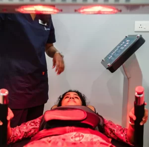
Maana Health provides physiotherapy management for ankylosing spondylitis across five clinics in Kerala — Kochi, Calicut, Perinthalmanna, Aluva, and Trivandrum. If you have chronic inflammatory back pain and have not been assessed for AS, book a free assessment today.